NEWS
Studierende etablieren bewegliches Abdomenphantom für Testzwecke unter MR-Bildgebung
Bis ein Medizinprodukt am Markt verfügbar ist, vergeht viel Entwicklungszeit, denn es müssen unter anderem genügend Testergebnisse zur Sicherheit und Leistung des Produktes gesammelt werden. Dazu sind je nach Klassifikation und Risikoeinschätzung des Medizinproduktes umfangreiche Studien und Tests an zum Beispiel Tieren, Tierkadavern, Gewebeproben oder Phantomen vorgeschrieben. Jedoch sind hierbei nur begrenzt die Gegebenheiten wie im menschlichen Körper nachstellbar, wie beispielsweise die Bewegungen bei der Atmung. Kommerziell erwerbbare Phantome sind zudem kostspielig. Die im Forschungscampus STIMULATE angesiedelte Forschungsgruppe um den Doktoranden Ivan Fomin hat ein MR-kompatibles Bewegungsphantom konzipiert, welches den Bereich des Abdomens (die obere Bauchregion) und die Organverschiebung durch die Atmung darstellt. Das Phantom soll zur Erprobung von Medizinprodukten und Prototypen im Bereich bildgestützter minimal-invasiver Interventionen dienen.
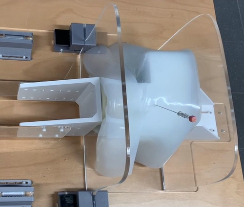
Die Arbeit richtete sich hierbei vor allem auf zwei Bereiche – die Konzeptionierung und Fertigung eines Abdomenphantoms und die Entwicklung einer Motoreinheit zur Simulation der Bewegung der Lungen und des Zwerchfells bei der Atmung. Die Konzeptionierung und Materialauswahl für das Phantom erörterte und erprobte die Medizintechnikstudentin Katja Engel im Rahmen ihrer Bachelorarbeit. Die Anforderungen an das Phantom sind zum einen die bildgebenden Eigenschaften, sodass es sich im MRT (Magnetresonanztomographen) menschlichem Bauchgewebe annähert, zum anderen die elastischen Eigenschaften, um ein reversibles Eindrücken mit dem Motor und ein realistisches Einstechverhalten mit zum Beispiel einer Ablationsnadel zu gewährleisten. Ein erstes Proof-of-Konzept-Phantom aus Polyvinylalkohol-Kryogel (PVA-K) wurde zusammen mit der von Ivan Fomin entwickelten Motoreinheit und mit Hilfe des Elektrotechnikstudenten Anton Schlünz erprobt. Eine eingeführte Nadel im Phantom zeigte bereits ein ähnliches Bewegungsmuster wie dies in einem Patienten aufgrund der Atmung zu beobachten ist (siehe Video-Links unten).
Aktuell wird die Weiterentwicklung des Motors und die Optimierung der Phantomeigenschaften in Bezug auf die Darstellung der Bauchorgane vorangetrieben. Weitere Schwerpunkte sind die realitätsgetreue Simulation der Atemzyklen und die Validierung der erzeugten Atembewegung.
Video 1: Nadelbewegung im Phantom
Video 2: Nadelbewegung im Patienten

Students establish movable abdominal phantom for testing under MR imaging
A lot of development time elapses before a medical device is available on the market because, among other things, sufficient test results must be collected on the safety and performance of the product. For this purpose, depending on the classification and risk assessment of the medical device, extensive studies and tests on, for example, animals, animal cadavers, tissue samples or phantoms are prescribed. However, the same conditions as in the human body can only be reproduced to a limited extent, such as the movements during respiration. Commercially available phantoms are also expensive. The research group around the PhD student Ivan Fomin, based in the research campus STIMULATE, has designed an MR-compatible motion phantom that represents the abdomen (the upper stomach region) and organ displacement due to respiration. The phantom will be used to test medical devices and prototypes in the field of image-guided minimally invasive interventions.
The work here was directed primarily in two areas - the conceptual design and fabrication of an abdominal phantom and the development of a motor unit to simulate the movement of the lungs and diaphragm during respiration. The conceptual design and material selection for the phantom was selected and tested by Katja Engel, a medical technology student, as part of her bachelor's thesis. The requirements for the phantom are on the one hand the imaging properties, so that it approximates human abdominal tissue in the MRI (magnetic resonance imaging), and on the other hand the elastic properties, in order to ensure reversible indentation with the motor and realistic piercing behavior with, for example, an ablation needle. A first proof-of-concept phantom made of polyvinyl alcohol cryogel (PVA-K) was tested together with the motor unit developed by Ivan Fomin and with the help of electrical engineering student Anton Schlünz. An inserted needle in the phantom already showed a movement pattern similar to that observed in a patient due to respiration (see video links below).
Currently, the further development of the motor and the optimization of the phantom properties with regard to the representation of the abdominal organs is being pursued. Another focus is the realistic simulation of the respiratory cycles and the validation of the generated respiratory motion.