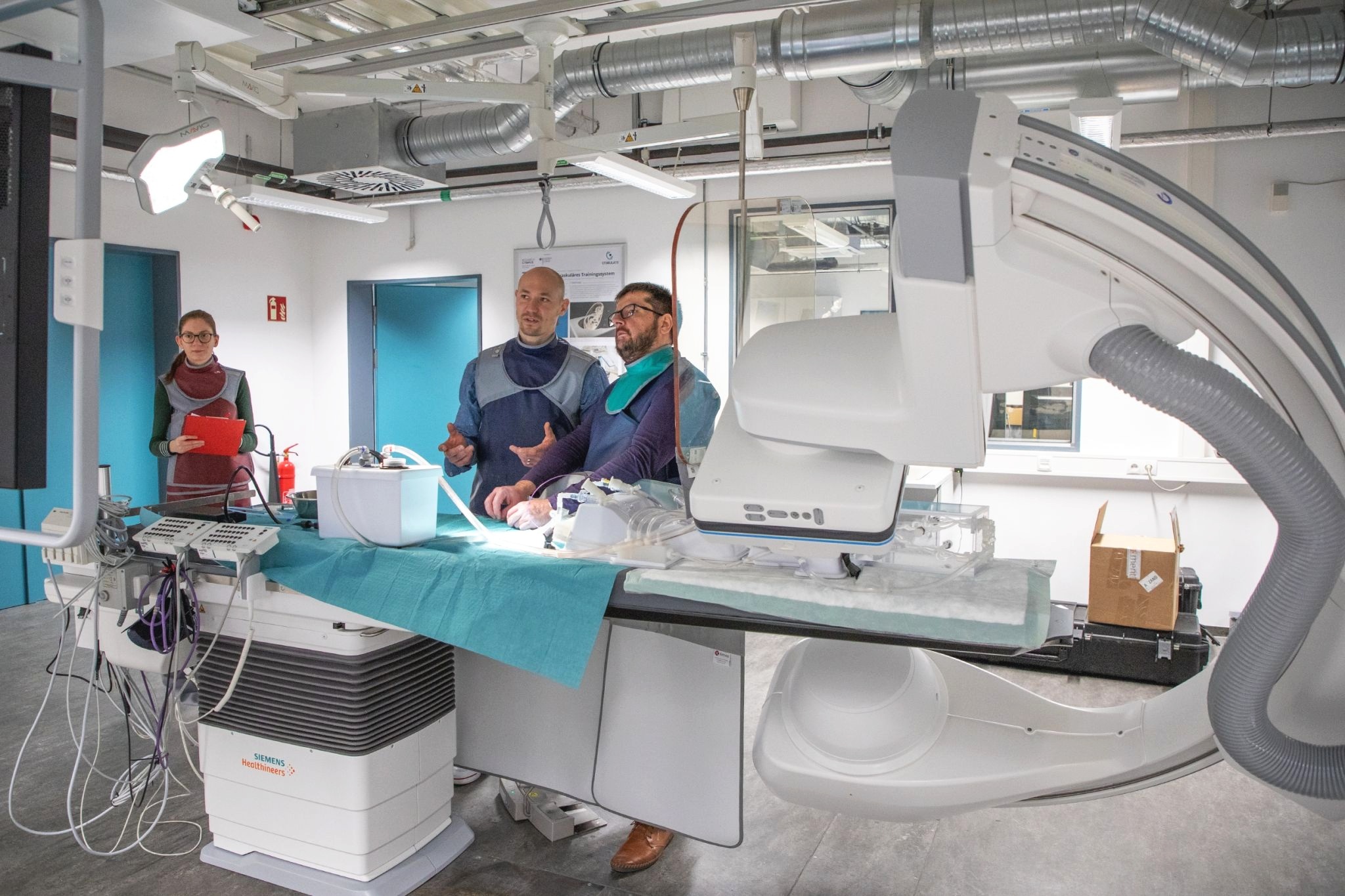
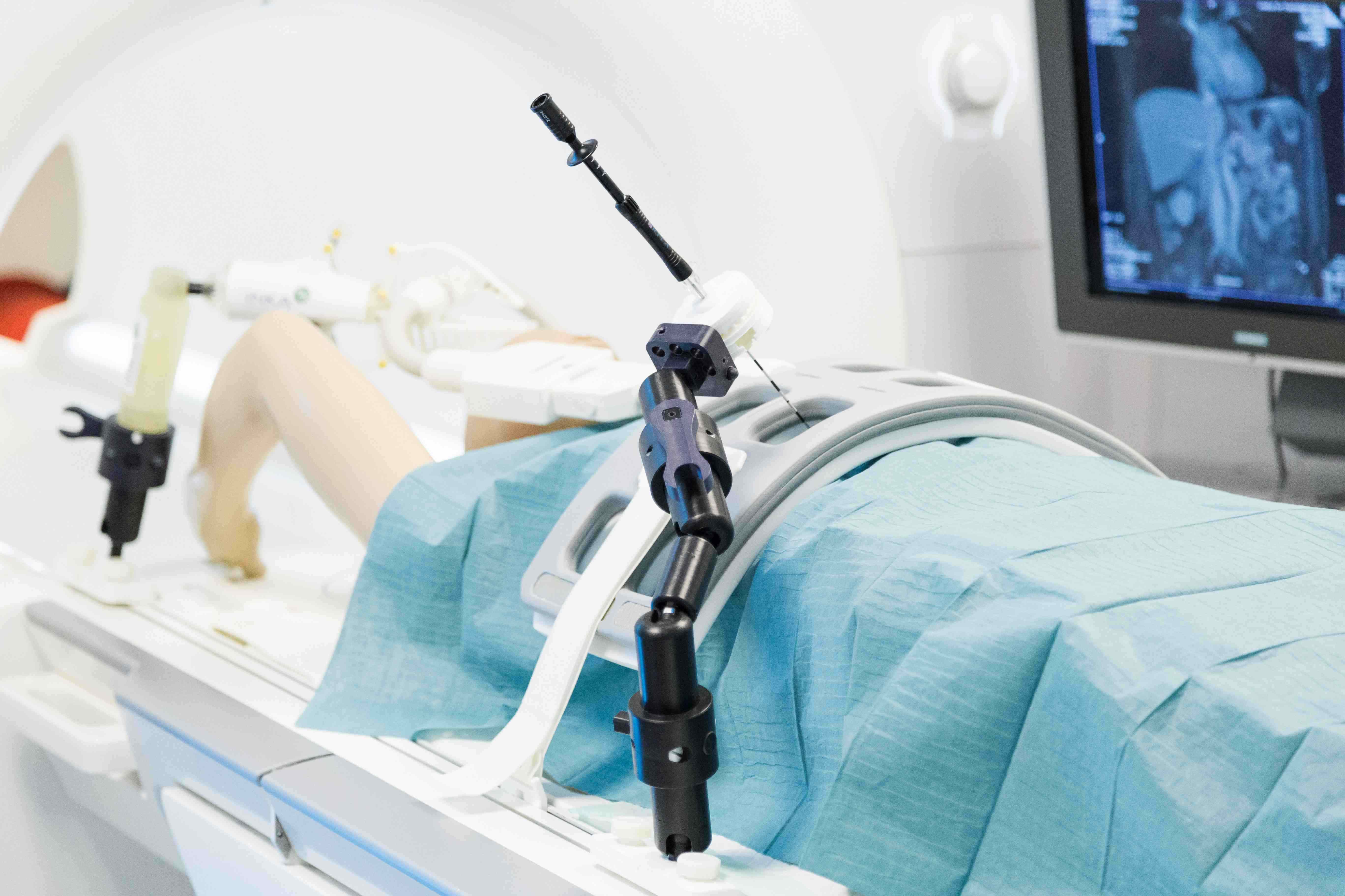
by Vanessa M. Swiatek, Belal Neyazi, Jorge A. Roa, Mario Zanaty, Edgar A. Samaniego, Daizo Ishii, Yongjun Lu, I. Erol Sandalcioglu, Sylvia Saalfeld, Philipp Berg, David M. Hasan
Abstract:
BACKGROUND: High-resolution vessel wall imaging plays an increasingly important role in assessing the risk of aneurysm rupture. OBJECTIVE: To introduce an approach toward the validation of the wall enhancement as a direct surrogate parameter for aneurysm stability. METHODS: A total of 19 patients harboring 22 incidental intracranial aneurysms were enrolled in this study. The aneurysms were dichotomized according to their aneurysm-to-pituitary stalk contrast ratio using a cutoff value of 0.5 (nonenhancing \textless 0.5; enhancing ≥ 0.5). We evaluated the association of aneurysm wall enhancement with morphological characteristics, hemodynamic features, and inflammatory chemokines directly measured inside the aneurysm. RESULTS: Differences in plasma concentration of chemokines and inflammatory molecules, morphological, and hemodynamic parameters were analyzed using the Welch test or Mann-Whitney U test. The concentration ΔIL-10 in the lumen of intracranial aneurysms with low wall enhancement was significantly increased compared to aneurysms with strong aneurysm wall enhancement (P = .014). The analysis of morphological and hemodynamic parameters showed significantly increased values for aneurysm volume (P = .03), aneurysm area (P = .044), maximal diameter (P = .049), and nonsphericity index (P = .021) for intracranial aneurysms with strong aneurysm wall enhancement. None of the hemodynamic parameters reached statistical significance; however, the total viscous shear force computed over the region of low wall shear stress showed a strong tendency toward significance (P = .053). CONCLUSION: Aneurysmal wall enhancement shows strong associations with decreased intrasaccular IL-10 and established morphological indicators of aneurysm instability.
Reference:
Aneurysm Wall Enhancement Is Associated With Decreased Intrasaccular IL-10 and Morphological Features of Instability. (Vanessa M. Swiatek, Belal Neyazi, Jorge A. Roa, Mario Zanaty, Edgar A. Samaniego, Daizo Ishii, Yongjun Lu, I. Erol Sandalcioglu, Sylvia Saalfeld, Philipp Berg, David M. Hasan), In Neurosurgery, volume 89, 2021.
Bibtex Entry:
@article{swiatek_aneurysm_2021,
	title = {Aneurysm {Wall} {Enhancement} {Is} {Associated} {With} {Decreased} {Intrasaccular} {IL}-10 and  {Morphological} {Features} of {Instability}.},
	volume = {89},
	copyright = {© Congress of Neurological Surgeons 2021.},
	issn = {1524-4040 0148-396X 0148-396X},
	doi = {10.1093/neuros/nyab249},
	abstract = {BACKGROUND: High-resolution vessel wall imaging plays an increasingly important role  in assessing the risk of aneurysm rupture. OBJECTIVE: To introduce an approach  toward the validation of the wall enhancement as a direct surrogate parameter for  aneurysm stability. METHODS: A total of 19 patients harboring 22 incidental  intracranial aneurysms were enrolled in this study. The aneurysms were dichotomized  according to their aneurysm-to-pituitary stalk contrast ratio using a cutoff value  of 0.5 (nonenhancing {\textless} 0.5; enhancing ≥ 0.5). We evaluated the association of  aneurysm wall enhancement with morphological characteristics, hemodynamic features,  and inflammatory chemokines directly measured inside the aneurysm. RESULTS:  Differences in plasma concentration of chemokines and inflammatory molecules,  morphological, and hemodynamic parameters were analyzed using the Welch test or  Mann-Whitney U test. The concentration ΔIL-10 in the lumen of intracranial aneurysms  with low wall enhancement was significantly increased compared to aneurysms with  strong aneurysm wall enhancement (P = .014). The analysis of morphological and  hemodynamic parameters showed significantly increased values for aneurysm volume  (P = .03), aneurysm area (P = .044), maximal diameter (P = .049), and nonsphericity  index (P = .021) for intracranial aneurysms with strong aneurysm wall enhancement.  None of the hemodynamic parameters reached statistical significance; however, the  total viscous shear force computed over the region of low wall shear stress showed a  strong tendency toward significance (P = .053). CONCLUSION: Aneurysmal wall  enhancement shows strong associations with decreased intrasaccular IL-10 and  established morphological indicators of aneurysm instability.},
	language = {eng},
	number = {4},
	journal = {Neurosurgery},
	author = {Swiatek, Vanessa M. and Neyazi, Belal and Roa, Jorge A. and Zanaty, Mario and Samaniego, Edgar A. and Ishii, Daizo and Lu, Yongjun and Sandalcioglu, I. Erol and Saalfeld, Sylvia and Berg, Philipp and Hasan, David M.},
	month = sep,
	year = {2021},
	pmid = {34245147},
	pmcid = {PMC8578742},
	keywords = {*Hemodynamics, *Inflammation, *Intracranial Aneurysm, *Magnetic Resonance Imaging, *Morphology, *Wall enhancement},
	pages = {664--671}
}